| Catalog Number | Size | Price | Quantity |
|---|---|---|---|
| FLD0703 | 0.5 mL | ¥329.0 | |
| FLD0703-1 | 1 mL | ¥589.0 |

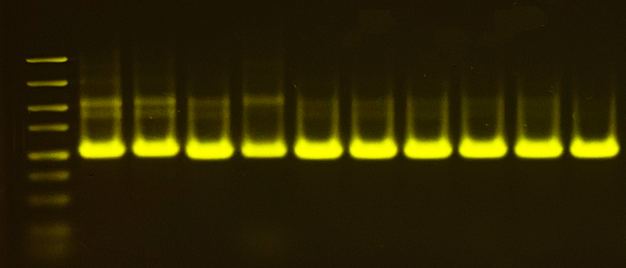
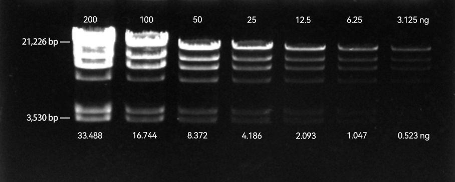
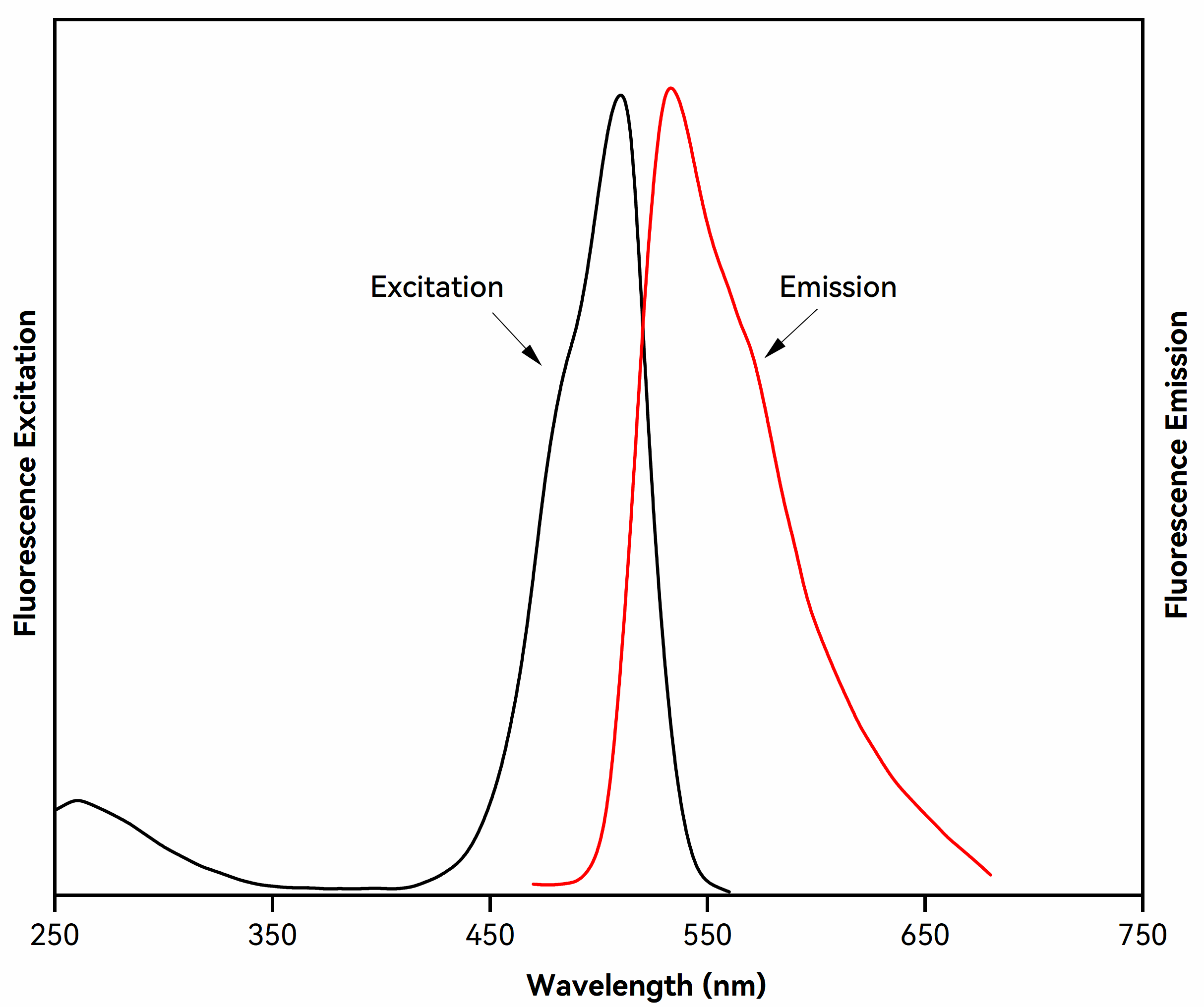
GelViewer is a sensitive, stable, low toxicity fluorescent nucleic acid dye for staining dsDNA, ssDNA and RNA in agarose or polyacrylamide gels. It is a safer and more effective alternative to ethidium bromide (EtBr). GelViewer provides excellent band visualization under both UV transilluminator and blue-light imagers, such as blue LED lightboxes. Using blue light helps prevent DNA damage and protect users from UV exposure. GelViewer support both in-gel staining (precast method) and post-staining, and is fully compatible with downstream applications, including gel purification, restriction digest, sequencing, and cloning.
| Issue | Solution |
|---|---|
| Smearing or trailing of DNA bands in pre-stained gels | 1. Reduce the DNA loading amount to 1/2 or 1/3 of the original. GelViewer is significantly more sensitive than ethidium bromide (EtBr), and overloading may cause overexposure or smearing of bands. |
| 2. Consider switching to post-staining instead of pre-staining. Post-staining minimizes interference with DNA migration and improves imaging quality. | |
| 3. For high molecular weight DNA, use a lower percentage agarose gel. Smearing is more common with large DNA fragments. | |
| 4. Use TBE buffer instead of TAE. TBE provides stronger buffering capacity and is better suited for high-resolution electrophoresis. | |
| 5. Avoid using loading buffers containing SDS, as SDS can disrupt dye–DNA binding and alter migration patterns. If SDS is required, post-staining is strongly recommended for optimal results. | |
| Altered DNA migration in pre-stained gels | 1. Reduce the DNA loading amount to 1/2 or 1/3 of the original. |
| 2. Lower the dye concentration; a 0.5× working concentration is recommended for in-gel staining. | |
| 3. Use post-staining instead of pre-staining to minimize interference with DNA migration and improve band resolution and imaging quality. | |
| Reduced fluorescence intensity | 1. The dye may have precipitated. Warm the solution at 40–50 °C for 1–2 minutes and vortex thoroughly to redissolve. |
| 2. Store the dye at room temperature and protect it from light to prevent precipitation caused by low temperatures. |